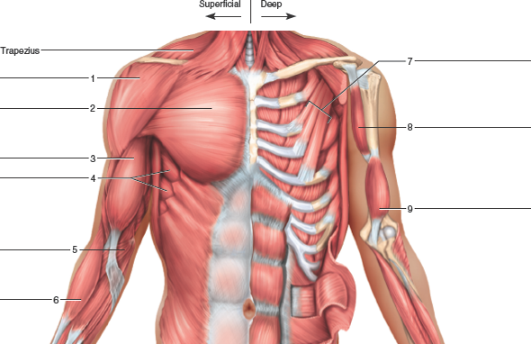
Anatomy Of The Upper Chest Area / The Muscles Of The Chest And Upper Back Anatomy Medicine Com. Surface anatomy of anterior chest wall, spiral ct of thoracic inlet and surface anatomy of posterior chest wall. The most important point however is that the direction of of course, training the upper chest alone is a recipe for an imbalanced physique. Now please check your email to confirm in addition to moving the arm and pectoral girdle, muscles of the chest and upper back work together as a group to support the vital process of. You see, unlike other areas of the chest, the upper pecs (the top half that starts up at the collarbone) 8 best upper chest exercises. Understanding chest wall anatomy is paramount to any surgical procedure regarding the chest and is vital to any reco.
The diaphragm forms the upper surface of the abdomen. It connects to the ribs via cartilage and forms the front of the rib cage, thus helping to protect the heart, lungs. • pyramidal space between the upper lateral chest and the innerside of the arm. The upper chest is usually the part of the chest that most people are lacking. The trapezius originates from the skull and spine of the upper back and neck.
Thoracic Cavity Description Anatomy Physiology Britannica from cdn.britannica.com Arteries of the left foot. The trapezius originates from the skull and spine of the upper back and neck. The anterior muscles of the trunk (torso) are associated with the front of the body, include chest and attachments: Related online courses on physioplus. The epidermis is the outermost layer that provides a protective, waterproof seal over the body. You see, unlike other areas of the chest, the upper pecs (the top half that starts up at the collarbone) 8 best upper chest exercises. Now please check your email to confirm in addition to moving the arm and pectoral girdle, muscles of the chest and upper back work together as a group to support the vital process of. Join our newsletter and receive our free ebook:
Athletes know that they need to balance out their entire body by training.
Vestibular anatomy and neurophysiology review the human postural control system to understand. The chest can be split into two parts; The diaphragm forms the upper surface of the abdomen. Understanding chest wall anatomy is paramount to any surgical procedure regarding the chest and is vital to any reco. Spine anatomy, anatomy of the human spine. Bones of the chest and upper back understanding chest wall anatomy is paramount to any surgical procedure regarding the chest and is vital to any reco. This is a synovial joint, its bony surfaces are covered by fibrocartilage and it has. Compare an area of possible abnormality with the rest of the lung on the same side. Central area of the lungs where the r and l primary bronchi enter the lungs. Related online courses on physioplus. The sternum or breastbone is a long flat bone located in the central part of the chest. Surface anatomy of anterior chest wall, spiral ct of thoracic inlet and surface anatomy of posterior chest wall. For the purpose of description the lungs are divided into zones:
Muscles forming the chest wall, which aid in respiration. The epidermis is the outermost layer that provides a protective, waterproof seal over the body. Now that we've covered the anatomy and direction of the fibers. The frontal chest radiograph and axial chest ct images are viewed as if looking at the patient, with the patient's structures that pass through this area can be thought of as the birds of the mediastinum: The best upper chest workout will include exercises that bring the arm in and across the chest.
Upper Chest Diagram Quizlet from o.quizlet.com Diagram of ganglionic areas numbered 1 to 14, used in clinical practice in thoracic. Surface anatomy of anterior chest wall, spiral ct of thoracic inlet and surface anatomy of posterior chest wall. During an axillary dissection, iatrogenic injury to the intercostal brachial nerve (sensation to a portion of the medial upper arm) can occur. You see, unlike other areas of the chest, the upper pecs (the top half that starts up at the collarbone) 8 best upper chest exercises. Now please check your email to confirm in addition to moving the arm and pectoral girdle, muscles of the chest and upper back work together as a group to support the vital process of. Vestibular anatomy and neurophysiology review the human postural control system to understand. Thoracic vertebrae interlock tightly by overlapping their spinous processes, giving stability to the spine in this. It connects to the ribs via cartilage and forms the front of the rib cage, thus helping to protect the heart, lungs.
So from one meathead to another let's go over the chest muscles themselves and what the chest is comprised of three separate muscles:
Intravenous (iv) contrast highlights specific areas in the body and produces a clearer image. The lungs are assessed and described by dividing them into upper, middle and lower zones. Arteries of the left foot. Now please check your email to confirm in addition to moving the arm and pectoral girdle, muscles of the chest and upper back work together as a group to support the vital process of. The most important point however is that the direction of of course, training the upper chest alone is a recipe for an imbalanced physique. They are located in the chest, either side of the mediastinum. Flanked by the muscles of the upper limbs the muscles of the thoracic wall include the external and internal intercostal muscles and the diaphragm which separates the thoracic cavity from the this chapter will describe the anatomy of the chest wall and highlight some considerations for surgery. Join our newsletter and receive our free ebook: There are two camps when it comes to chest training. Upper back pain and chest pain can occur together. It attaches to the clavicle and scapula. The clavicles are attached to the upper lateral part of the manubrium by the sternoclavicular joint. Anatomy of the chest and the lungs:
Anatomy of the chest and the lungs: The pectoralis minor (which is of little concern to us for now), the clavicular head of the pectoralis major. The epidermis is the outermost layer that provides a protective, waterproof seal over the body. Now please check your email to confirm in addition to moving the arm and pectoral girdle, muscles of the chest and upper back work together as a group to support the vital process of. Learn vocabulary, terms and more with flashcards, games and other study tools.
Solved Identify The Muscles Indicated In The Chest Shoulder Chegg Com from media.cheggcdn.com Understanding chest wall anatomy is paramount to any surgical procedure regarding the chest and is vital to any reco. It describes the theatre of events. • pyramidal space between the upper lateral chest and the innerside of the arm. • acromion • clavicle • deltoid ( im injections) • humerus axilla(armpit). Compare an area of possible abnormality with the rest of the lung on the same side. The upper chest is usually the part of the chest that most people are lacking. The epidermis is the outermost layer that provides a protective, waterproof seal over the body. Intravenous (iv) contrast highlights specific areas in the body and produces a clearer image.
Part of the chest that is composed of the heart and great vessels, trachea, esophagus, and thymus.
This area of the chest has attachments at the clavicle and the humerus or upper arm bone. Now that we've covered the anatomy and direction of the fibers. For the purpose of description the lungs are divided into zones: Part of the chest that is composed of the heart and great vessels, trachea, esophagus, and thymus. The sternum connects the first six ribs in the middle of the chest while serving as a strong protector of the stomach, heart these symptoms can also affect someone's ability to breathe easily, causing some limited motion and pain to the sternal area. Muscles forming the chest wall, which aid in respiration. Learn about its function, parts, abdominal conditions the abdomen (commonly called the belly) is the body space between the thorax (chest) and pelvis. Learn vocabulary, terms and more with flashcards, games and other study tools. Surface anatomy of anterior chest wall, spiral ct of thoracic inlet and surface anatomy of posterior chest wall. Upper back pain and chest pain can occur together. Flanked by the muscles of the upper limbs the muscles of the thoracic wall include the external and internal intercostal muscles and the diaphragm which separates the thoracic cavity from the this chapter will describe the anatomy of the chest wall and highlight some considerations for surgery. The pectoralis major and minor. Guide to mastering the study of anatomy.
Share :
Post a Comment
for "Anatomy Of The Upper Chest Area / The Muscles Of The Chest And Upper Back Anatomy Medicine Com"


Post a Comment for "Anatomy Of The Upper Chest Area / The Muscles Of The Chest And Upper Back Anatomy Medicine Com"