Musclular System Labeled Back ~ 16 563 Muscular System Stock Photos Images Download Muscular System Pictures On Depositphotos. Some conditions, including spinal stenosis and scoliosis, cause structural problems in the back, leading to pain and limited mobility. Muscles, connected to bones or internal organs and blood vessels, are in charge for movement. Each of these muscles is a discrete organ constructed of skeletal muscle tissue, blood vessels, tendons, and nerves. 5 out of 5 stars (1,528) 1,528 reviews Muscles vary greatly in their shape and size, depending on their function.
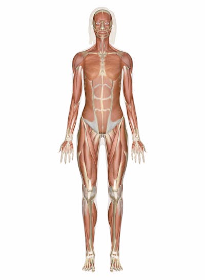
Human anatomy for muscle, reproductive, and skeleton. Muscles, connected to bones or internal organs and blood vessels, are in charge for movement. Their main function is contractibility. Almost every movement in the body is the outcome of muscle contraction. Unlike other organ systems, the muscular system is divided into different types of tissues, which are incorporated into various organs in the body.
Muscular System Vector Hd Stock Images Shutterstock from image.shutterstock.com Muscles vary greatly in their shape and size, depe Human anatomy for muscle, reproductive, and skeleton. The muscular systems in vertebrates are controlled through the nervous system although some. In addition, the axial skeleton that runs vertically through the back protects the spinal cord, which innervates almost all the muscles in the body. The muscles of the abdomen, lower back, and pelvis are separated from those of the chest by the muscular wall of the diaphragm, the critical breathing muscle. Each of these muscles is a discrete organ constructed of skeletal muscle tissue, blood vessels, tendons, and nerves. To see a muscular system picture from the anterior (front) view click here. These structures work together to support the body, enable a range of movements, and send messages from the brain to the.
Lying exposed between the protective bones of the superiorly located ribs and the inferiorly located pelvic girdle, the muscles of this region play a critical role in protecting the.
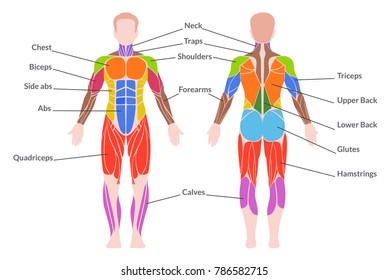
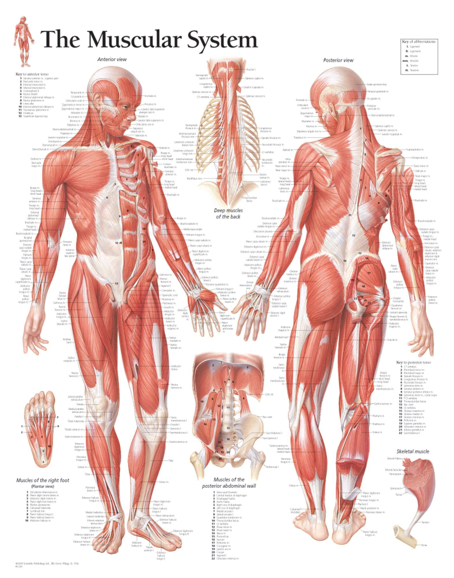
Human muscular system anatomy vintage poster torso muscles print doctor office decor surgeon gift physical therapist gift medical art homelibraryart. Their main function is contractibility. The muscular system's main function is to allow movement. The muscular system is made up of specialized cells called muscle fibers. This labeled human muscular system chart illustrates the major muscle groups in the back (posterior) view and the front (anterior) view. Without muscle, humans could not live. We think this is the most useful anatomy picture that you need. Superficial back muscles, intermediate back muscles and intrinsic back muscles.the intrinsic muscles are named as such because their embryological development begins in the back, oppose to the superficial and intermediate back muscles which develop elsewhere and are therefore classed as extrinsic muscles. To access the teachmeanatomy 3d model, you must be a registered subscriber. Muscular system, human muscles art, physical therapist gift, med student gift, back muscle, massage therapist office enorasis. The muscles of the back can be arranged into 3 categories based on their location: Muscle tendons in the knee joint and the shoulder joint are crucial in stabilization. In addition, the axial skeleton that runs vertically through the back protects the spinal cord, which innervates almost all the muscles in the body.
The most realistic hd human male muscular system modeled in zbrush and was extracted from our hd male complete human 3d anatomy model. This labeled human muscular system chart illustrates the major muscle groups in the back (posterior) view and the front (anterior) view. The muscle fibers' highly specialized structure enables the muscles to relax and contract to produce movement. Their main function is contractibility. Leg muscle anatomy (front view.
The Female Muscular System Scientific Publishing from www.scientificpublishing.com The primary job of muscle is to move the bones of the skeleton, but muscles also enable the heart to beat and constitute the walls of other important hollow. The muscles of the back can be arranged into 3 categories based on their location: Human muscular system anatomy vintage poster torso muscles print doctor office decor surgeon gift physical therapist gift medical art homelibraryart. Muscles vary greatly in their shape and size, depending on their function. Superficial back muscles, intermediate back muscles and intrinsic back muscles.the intrinsic muscles are named as such because their embryological development begins in the back, oppose to the superficial and intermediate back muscles which develop elsewhere and are therefore classed as extrinsic muscles. This labeled human muscular system chart illustrates the major muscle groups in the back (posterior) view and the front (anterior) view. Human muscle system muscles of the back. The core muscles are those in the.
Each of these muscles is a discrete organ constructed of skeletal muscle tissue, blood vessels, tendons, and nerves.
The muscle fibers' highly specialized structure enables the muscles to relax and contract to produce movement. The muscles of the back can be arranged into 3 categories based on their location: Human muscle system muscles of the back. Some conditions, including spinal stenosis and scoliosis, cause structural problems in the back, leading to pain and limited mobility. The back consists of the spine, spinal cord, muscles, ligaments, and nerves. Back pain in human body. Anatomynote.com found anatomy of back muscles diagram from plenty of anatomical pictures on the internet. This muscular system diagram shows the major muscle groups from the back or posterior view. More specifically, this beautifully illustrated anatomy chart includes head and neck, thorax, multiple abdominal view, and frontal views of each muscular extremity of the human body. Each of your muscles is made up of thousands of thin, long, cylindrical cells called muscle fibers. Attached to the bones of the skeletal system are about 700 named muscles that make up roughly half of a person's body weight. For more anatomy content please follow us and visit our website: The back contains the origins of many of the muscles that are involved in the movement of the neck and shoulders.
Muscles, connected to bones or internal organs and blood vessels, are in charge for movement. The back consists of the spine, spinal cord, muscles, ligaments, and nerves. Learn vocabulary, terms, and more with flashcards, games, and other study tools. This type of muscle attaches to the skeleton and moves the limbs and body of an organism. Leg muscle anatomy (front view.
Muscular System Muscles Of The Human Body from innerbody.imgix.net Human musculature bodybuilding infographic muscular system vector human anatomy back muscle anatomy bicep male muscular anatomy human body anatomy female female anatomy muscle hamstrings muscle. Muscles vary greatly in their shape. To access the teachmeanatomy 3d model, you must be a registered subscriber. Several types of cancer affect the musculoskeletal system, including bone cancer. This muscular system diagram shows the major muscle groups from the back or posterior view. The muscular system is responsible for the movement of the human body. The muscular systems in vertebrates are controlled through the nervous system although some. Muscles vary greatly in their shape and size, depe
The back contains the origins of many of the muscles that are involved in the movement of the neck and shoulders.
The core muscles are those in the. Human musculature bodybuilding infographic muscular system vector human anatomy back muscle anatomy bicep male muscular anatomy human body anatomy female female anatomy muscle hamstrings muscle. We think this is the most useful anatomy picture that you need. Muscles vary greatly in their shape. Anatomynote.com found anatomy of back muscles diagram from plenty of anatomical pictures on the internet. To access the teachmeanatomy 3d model, you must be a registered subscriber. To see a muscular system picture from the anterior (front) view click here. Top view of female internal organs on brown. The muscular system is responsible for the movement of the human body. More specifically, this beautifully illustrated anatomy chart includes head and neck, thorax, multiple abdominal view, and frontal views of each muscular extremity of the human body. Muscle tendons in the knee joint and the shoulder joint are crucial in stabilization. Human muscle system muscles of the back. Muscle tendons stretch over joints and contribute to joint stability.
Share :
Post a Comment
for "Musclular System Labeled Back ~ 16 563 Muscular System Stock Photos Images Download Muscular System Pictures On Depositphotos"

Post a Comment for "Musclular System Labeled Back ~ 16 563 Muscular System Stock Photos Images Download Muscular System Pictures On Depositphotos"